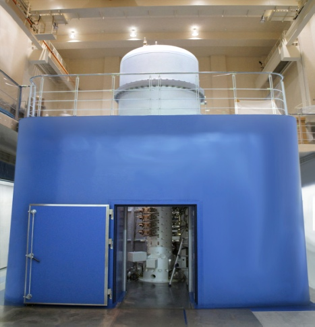
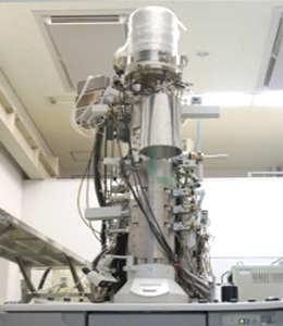
Isotope Microscope – IMS-1270 / CAMECA
Creative Research Institution SOUSEI, Hokkaido University
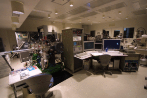
This instrument is a development of CAMECA’s ultra-high mass resolution Secondary Ionization Mass Spectrometer: IMS-1270 technology and enables imaging of the three-dimensional distribution of isotopes in a material. It has a spatial resolution of 300 nm in the lateral direction and 10 nm in the depth direction for isotopes with concentrations up to 1 ppb, and enables isotope ratio analysis with a relative error of 0.5% for isotopes with a 500-fold difference in concentration. More information, Details
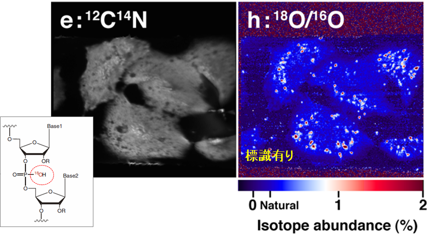
Isotope ratio imaging of the uptake of 18O isotope-labelled RNA into cultured cells. Hamasaki et al., Nucleic Acids Research (2013)
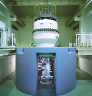
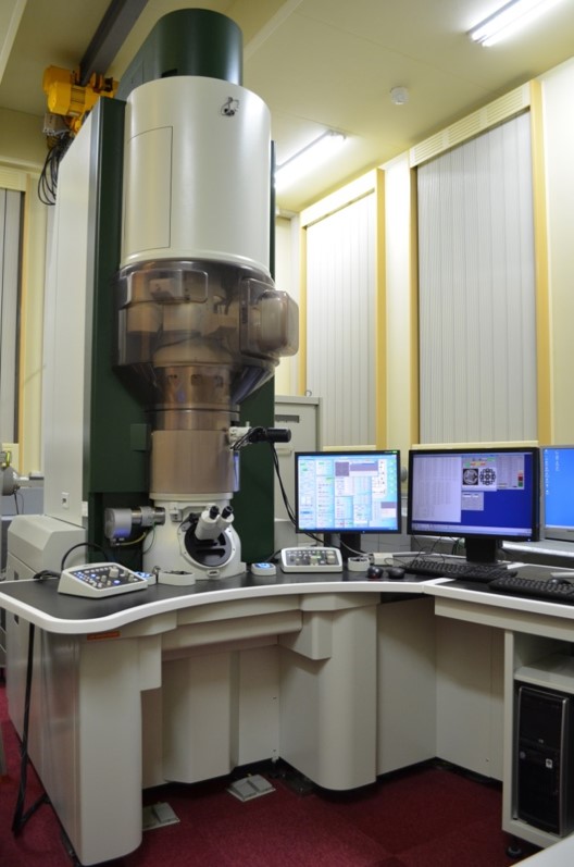
Next Generation Isotope Microscope – IMS-1280HR / CAMECA
Creative Research Institution SOUSEI, Hokkaido University
Enables ultra-sensitive mass analysis, especially in point analysis. Higher mass resolution than isotope microscope. More information Details
MALDI-IT-TOF Imaging Mass Microscope – iMScope beta / SHIMADZU
International Mass Imaging and Spatial Omics Center, Hamamatsu University School of Medicine
This instrument was developed by our laboratory and SHIMADZU with the support of “JST-SENTAN (Development of Advanced Measurement and Analysis Systems)” program. It enables high-resolution measurements with laser diameters of 5 µm or less. More information
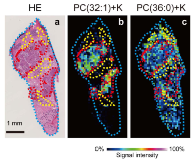
Observations on phosphatidylcholine (PC) in human breast cancer tissue. PC (32:1) was found to be more abundant in cancerous areas (red) and PC (36:0) in non-cancerous areas (yellow).
Ultra-high Resolution FT-ICR Mass Spectrometer – Solarix XR / Bruker Japan
International Mass Imaging and Spatial Omics Center, Hamamatsu University School of Medicine
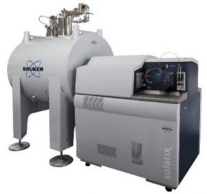
Fourier Transform Ion Cyclotron Resonance mass spectrometer enables ultra-sensitive analysis with super-resolution of up to 10 million FWHM. Imaging is possible with a spatial resolution of 10 µm. More information
Operations will carry out by institution’s specialist researchers.
DESI Quadrupole Time-of-Flight Mass Spectrometer – Xevo® G2-XS Qtof / Waters
International Mass Imaging and Spatial Omics Center, Hamamatsu University School of Medicine
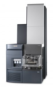
Imaging by soft ionization with Desorption ElectroSpray Ionization, without the need for matrix application or other pretreatment. Capable of measuring free fatty acids, for example. The Quadrupole-Time-of-Flight mass spectrometer allows high-resolution measurements and structure estimation by MS/MS measurements (product ion spectrometry). More information
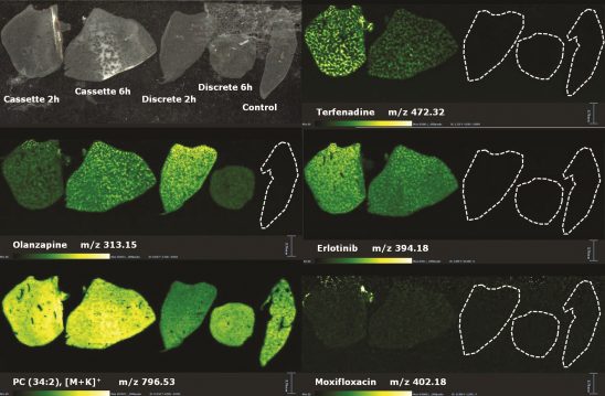
The distribution of drugs and phosphatidylcholine (PC) was visualised for mouse livers 2 hours and 6 hours after administration of multiple compounds.
DESI Mass Spectrometer – Xevo® TQ-XS / Waters
International Mass Imaging and Spatial Omics Center, Hamamatsu University School of Medicine
This equipment allows imaging using the same soft ionization technique as the DESI-Qtof. The triple quadrupole mass spectrometer enables highly selective and sensitive imaging by means of selected reaction monitoring (SRM). SRM, also known as MRM, is a generic measurement method for highly sensitive quantitative analysis by LC/MS. More information
As this equipment is located in an Radio Isotope controlled area, the researcher in charge will accompany you to operate the system when in use,
3D-SIM Super-resolution Imaging System – DeltaVision OMX / GE Healthcare
Natural Science Center for Basic Research and Development, Hiroshima University
3D-SIM is a fluorescence microscope that uses the moire fringe phenomenon caused by laser interference to achieve super-resolution. It has spatial resolution of up to 90 nm in the x and y axes and 220 nm in the z axis, enabling more detailed imaging and analysis of intracellular structures and microstructures than conventional fluorescence microscopes. And this system also enables high-speed image acquisition and can be adapted for live imaging. More information
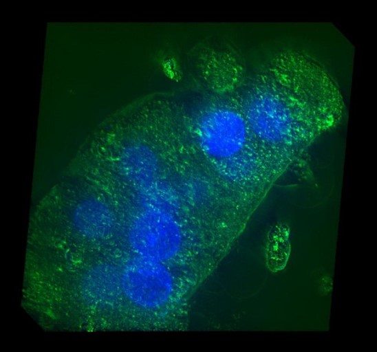
Three-dimensional cultured cells (primary rat hepatocytes) were measured and the cell population was observed. see also imaging with JSM-7800F
Thermal FE-SEM – JSM-7800F / JEOL
Natural Science Center for Basic Research and Development, Hiroshima University
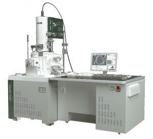
This Scanning Electron Microscope has high-performance electron optics. Secondary electrons maand reflected electrons are emitted from the sample. The secondary electrons mainly provide information on the surface irregularities of the sample, while the reflected electrons provide information on the atomic numbers of the sample’s constituent materials. In addition, elemental analysis can be performed from these reflected electrons using an energy-dispersive X-ray spectrometer (JED-2300). This instrument is also equipped with a cryotransfer system (ALTO 2500), which enables observation of water-containing or heat-sensitive samples. More information
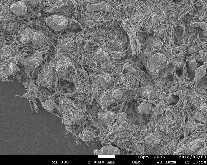
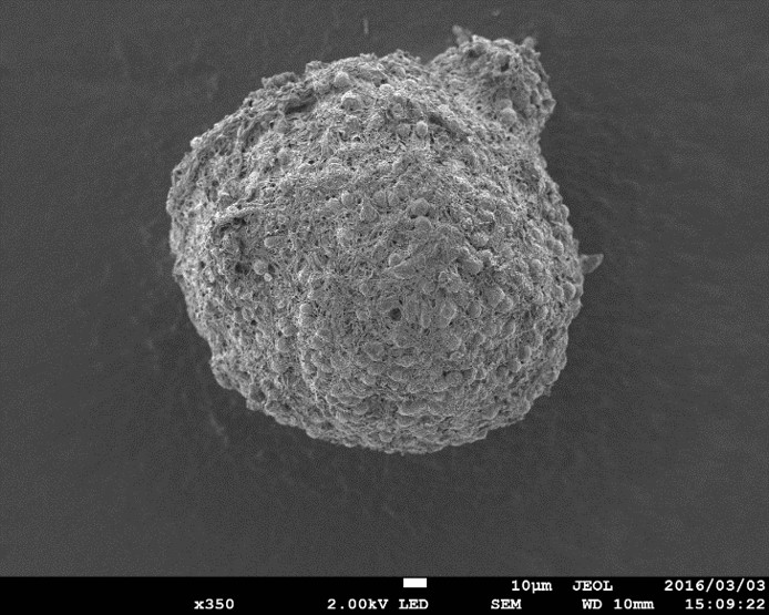
Spheroids (cell aggregate)in primary rat cerebral cortex cells. Combined with the results of the analysis by 3D-SIM , the actual three-dimensional structure of the spheroid was confirmed.
Confocal Laser Scanning Microscope – FV1000-D / OLYMPUS
Natural Science Center for Basic Research and Development, Hiroshima University
Confocal laser scanning microscopes allow samples to be observed with higher resolution than conventional fluorescence microscopes, It is also possible to construct a three-dimensional image of the sample by acquiring continuous tomographic images in the depth direction.
This is used for cell collection under a microscope, which is a pre-treatment for live single-cell mass spectrometry. More information
Imaging Mass Microscope System – iMScope / SHIMAZDU
Natural Science Center for Basic Research and Development, Hiroshima University
Molecular information up to a molecular weight of 3,000 can be obtained using the MALDI method for each micro-region on the order of µm for a sample section on a glass slide, while moving the measurement position.
The sample section on a glass slide is photographed using built-in microscope, and by superimposing the mass spectrum of each micro-region obtained on the image, the molecular distribution of the sample can be observed. More information
Atomic Resolution Holography Electron Microscope (1.2 MV)
Hitachi, Ltd. Research & Development Group
This cutting-edge 1.2 MV atomic resolution holography transmission electron microscope not only has high penetrability due to high acceleration, it also has outstanding performance in terms of spatial resolution with an aberration corrector for ultra-high-voltage TEMs. More information
Electron accelerating voltage: 1.2 MV
Ultra-high resolution (up to 43 pm)
Spherical aberration corrector
Electron Holography
Magnetic-field-free observation stage
Applies magnetic fields (up to 500mT)
Liquid Helium Cooling
* This device was developed with the assistance of the Japan Society for the Promotion of Science under the Funding Program for World-Leading Innovative R&D on Science and Technology (Cabinet Office) planned by the Council for Science, Technology and Innovation.
Ultra-high-voltage Holography Electron Microscope (1 MV)
Hitachi, Ltd. Research & Development Group
This 1MV holography TEM has a sample cooling mechanism for realizing stable temperatures from room temperature to extremely low temperatures (down to 5 K). It also has a mechanism that allows the applying of a magnetic field to the sample from any direction, for which it is being used in research on observations of flux quantum in superconductors. more
Electron accelerating voltage: 1 MV
High resolution (up to 120 pm)
Electron Holography
Magnetic-field-free observation stage
Applies magnetic fields (up to 500mT)
Liquid Helium Cooling
350 kV Holography TEM
Hitachi, Ltd. Research & Development Group
Electron accelerating voltage: 350 kV
High resolution (up to 200 pm)
Electron Holography
Applies magnetic fields (up to 100mT)
i-hitachi
300 kV Holography TEM – HF3300EH
Nanostructures Research Laboratory, Japan Fine Ceramics Center
Electric potential observation in semiconductors, magnetic flux observation in magnetic materials. More information
Acceleration voltages are available for 100, 200, 300 kV.
4 electron biprisms are installed.
High resolution (up to 240 pm)
Special TEM holders are available (Heating, Cooling, Biasing).
Applying magnetic fields (up to 120 mT)
300 kV Holography TEM
The Ultramicroscopy Research Center, Kyushu University
One of the most experienced in the world in metallic specimen research. More information
1. Combined electromagnetic field analysis synchronized with structural and compositional analysis.
2. Analysis of electron-irradiation-sensitive samples through the use of a wide range of acceleration voltage settings and the use of high-speed, high-sensitivity cameras.
Electron accelerating voltage: 300kV, 200kV, 100kV
High resolution (up to 200 pm)
Heating and cooling of samples
Applies magnetic fields (up to 50 mT)
TEM – ARM-200F / JEOL
Institute of Materials and Systems for Sustainability, Nagoya University
High-resolution TEM/STEM observation
Chemical analysis equipment for experienced users.
More information
Electron accelerating voltage: 200kV
Double aberration correction
Electron biprism
EDX / EELS Spectrometer
Backscattered electron detector
Combined Electron Spectroscopy Scanning Electron Microscope – JEM2100 / JEOL
Institute of Materials and Systems for Sustainability, Nagoya University
Chemical analysis of site-selective dopants by combined electron microspectroscopy
Material space mapping
3D analysis of crystal lattice defects.
More information
Electron accelerating voltage: 200kV
Thermionic-emission gun
EDX / WDX / EELS / CL spectrometer
Backscattered electron detector
Automatic Freeze Substitution System – EM AFS2 / Leica Microsystems
Creative Research Institution SOUSEI, Hokkaido University
This equipment can be used for sample preparation for isotope microscope. Progressive Lowering of Temperature (PLT) allows substitution and resin infiltration of chemically fixed specimens.
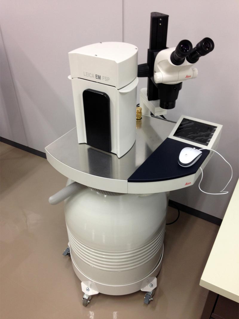
High Pressure Freezer – EM HPM100 / Leica Microsystems
Creative Research Institution SOUSEI, Hokkaido University
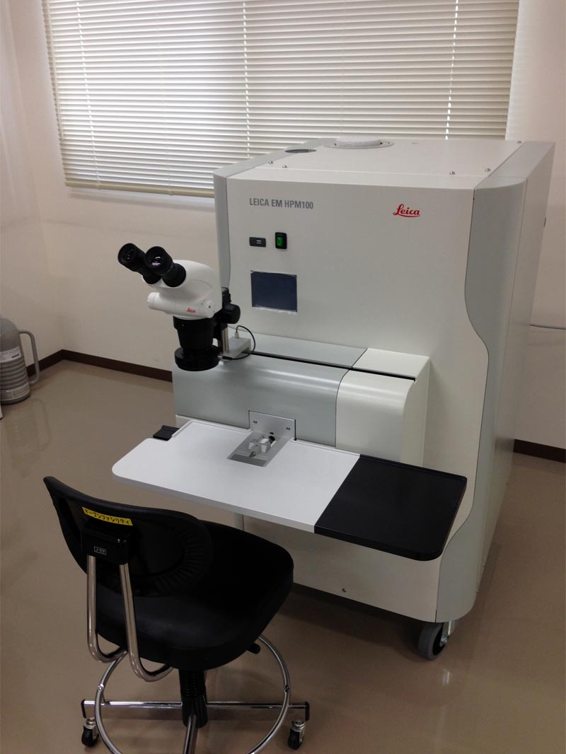
This equipment can be used for sample preparation for isotope microscope. The 2100 bar of high pressure applied to the sample during high pressure freezing using this equipment suppresses ice crystal formation and growth, while cryo immobilization immediately after pressurization prevents structural damage to the sample. More information
Ultramicrotome – EM UC7i / Leica Microsystems
Creative Research Institution SOUSEI, Hokkaido University
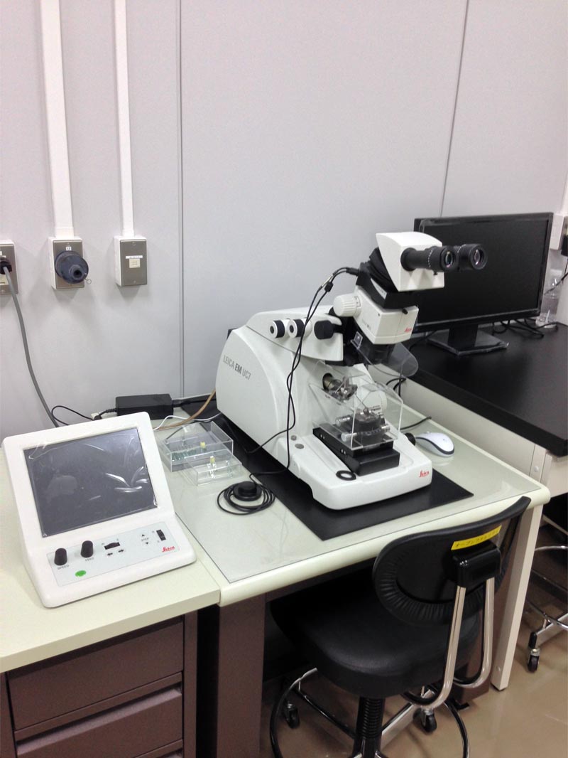
This equipment can be used for sample preparation for isotope microscope. High quality sectioning of specimens examination can be easier and more precise. More information
3D Laser Scanning Microscope System – VK-X200 / KEYENCE
Creative Research Institution SOUSEI, Hokkaido University
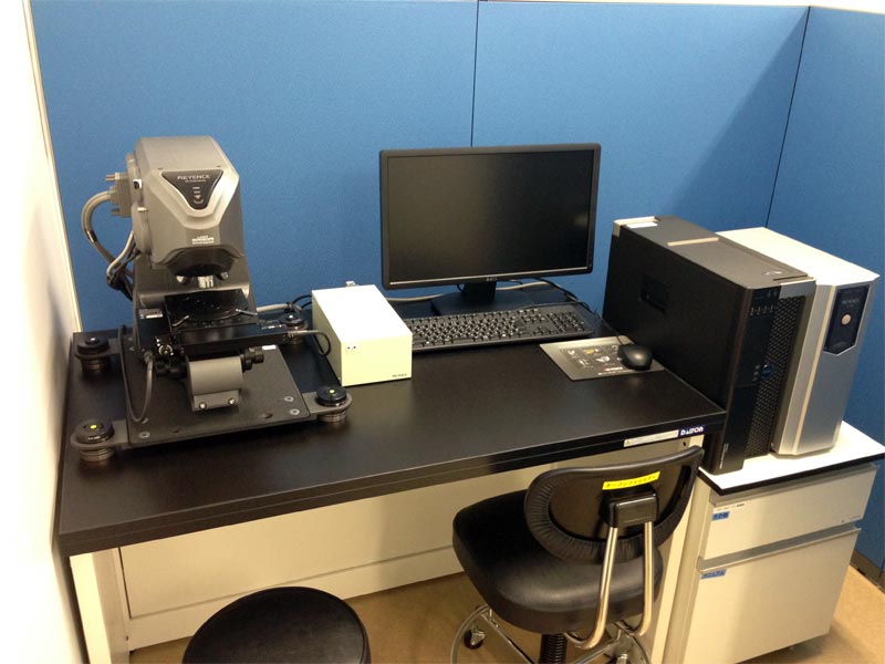
Easy operation to obtain high resolution focal image and gather height information (shape and roughness) from the sample. More information
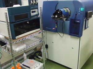
Liquid Chromatography-Mass Spectrometer – SYNAPT-G2 HDMS / Waters
International Mass Imaging and Spatial Omics Center, Hamamatsu University School of Medicine
LC-QTOF-MS system with electrospray ion source and ion mobility mass separation. Fast, high-resolution mass spectrum acquisition and MS/MS (product ion) analysis are possible with UHPLC. More information
Cryostat for Infection Prevention and Control – CM1950 / Leica Biosystems
International Mass Imaging and Spatial Omics Center, Hamamatsu University School of Medicine
Equipped with UV treatment and a nanosilver surface coating. More information
Matrix Vapor Deposition System – iMLayer / SHIMADU
International Mass Imaging and Spatial Omics Center, Hamamatsu University School of Medicine
Automatic matrix coating system for MALDI imaging samples.
Matrix Spraying System – TM-Sprayer / HTX Imaging
International Mass Imaging and Spatial Omics Center, Hamamatsu University School of Medicine
Automatic matrix coating system for MALDI imaging samples. More information
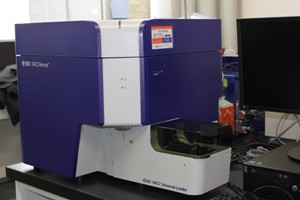
Cell Sorter – FACS Aria II SORP / Becton Dickinson
Natural Science Center for Basic Research and Development, Hiroshima University

In addition to the flow cytometry function, This equipment has a cell sorting function. More information
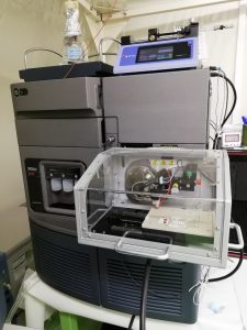
HPLC Mass Spectrometer – LCMS-8050 / SHIMAZDU
Natural Science Center for Basic Research and Development, Hiroshima University
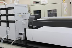
This system consists of a high-performance liquid chromatograph (Nexera X2) and a triple quadrupole mass spectrometer (LCMS-8050). ESI and APCI methods are available. More information
Cell Analyzer – LSRFortessa X-20 / Becton Dickinson
Natural Science Center for Basic Research and Development, Hiroshima University
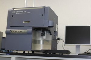
This Equipment is a flow cytometer equipped with a high-throughput sampler and is operationally compatible with FACSAria. More information
Cell Analyzer – FACSVerse / Becton Dickinson
Natural Science Center for Basic Research and Development, Hiroshima University
This analyzer enables multi-colour analysis, detecting eight fluorescent colours simultaneously. It is also equipped with a universal loader, which enables automated sampling of multiple specimens using microplates or tube racks. More information
Mass Spectrometer – TripleTOF 5600+ / AB SCIEX
Natural Science Center for Basic Research and Development, Hiroshima University
Q-TOF mass spectrometer capable of using the nanoESI, ESI and APCI methods. It can also be used in combination with the ion mobility separation system ‘SelexION’ for the separation of isobaric compounds. More information